Stapedectomy Surgery
Stapedectomy surgery is a procedure in which the innermost bone (stapes) of the three bones (the stapes, the incus, and the malleus) of the middle ear is removed and replaced with a prosthesis. Performed to improve the movement of sound to the inner ear, stapedectomy treats progressive hearing loss caused by otosclerosis, a condition in which spongy bone hardens around the base of the stapes.
A stapedectomy is an outpatient surgical procedure done under local or general anesthesia through the ear canal with an operating microscope. It involves removing the immobilized or fixed stapes bone and replacing it with a prosthetic device. The prosthetic device allows the bones of the middle ear to resume movement, which allows sound to properly reach the fluid in the inner ear and thereby improves or restores hearing.
Modern-day stapedectomies have been performed since 1956 with a success rate of 95 percent. In rare cases (about one percent of surgeries), the procedure may worsen hearing. When otosclerosis has affected both ears, the ears are operated on one at a time, with a four-to six-month time gap between surgeries.
Surgical Procedure
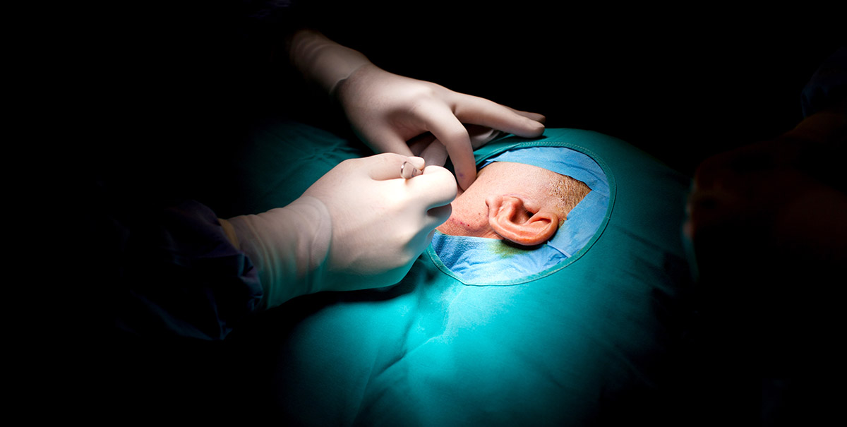
A stapedectomy is performed under general or local anesthesia in the operating room. The binocular operating microscope is used for high magnification of the very small structures in the middle ear such as the stapes footplate, which measures 2mm by 3mm.
The operation is performed through the ear canal with specialized microsurgical instruments. With general anesthesia, the surgery usually takes about an hour. After the tympanic membrane (also known as the eardrum) is elevated, the middle ear is entered. The middle bones of hearing are examined and their ability to move is noted. The third bone of hearing, the stapes, is examined for fixation and visible otosclerosis deposits. If these are present, otosclerosis is diagnosed. The upper portion of the stapes bone is then vaporized with a Laser. The distance between the fixed stapes footplate and the mobile incus (2nd bone used for hearing) is measured to select the properly sized prosthesis. Approximately 0.7mm hole in the fixed stapes footplate is created with the Laser. The prosthesis is then inserted into the footplate hole and then “welded” to the incus with the Laser. A small piece of tissue from the back of the ear lobule is draped around the prosthesis at the footplate hole to seal it. The prosthesis is checked for movement when the first two bones of hearing are moved to ensure that sound energy can be easily transmitted through the footplate to the inner ear. The eardrum is returned to its normal position and secured with ear canal packing.

What’s Going On At BSSNY
-

-

Regional neurosurgery practice can now accept major insurance plans
19 September, 2022 -

Functional neurosurgery brings relief to patients that had given up
29 December, 2021 -

Brain and Spine Surgeons of New York expands practice to Stamford
01 December, 2021