
Cervical Vertebrectomy
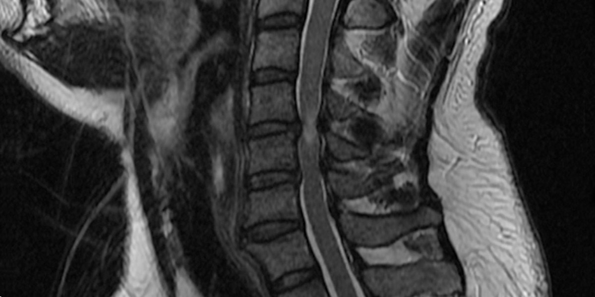
A cervical vertebrectomy is performed when degenerative changes in the cervical spine have resulted in the formation of bone spurs and/or herniated discs. When this happens, the spinal canal narrows, which can result in compressed (pinched) nerves and pain.
In addition to degenerative diseases such as stenosis, vertebrae that have been damaged by trauma, tumors, or deformities may also benefit from a cervical vertebrectomy.
The Procedure
After making a small incision in the patient’s neck, the surgeon will remove the vertebra as well as the disc spaces at either end in order to relieve the pressure on the spinal cord and nerves. In most cases, the surgeon will then perform a spinal fusion by filling the now-empty space with an implant. This implant fills the space and provides strength and stability.
There are several different types of implants your surgeon may use; which depends on the patient’s individual circumstances. These include:
- Bone graft – Taken from the patient’s own body or a bone bank, the graft is inserted into the empty space and fixed in place with titanium screws and plates. This kind of graft will eventually fuse with the patient’s body to provide long-term spinal stability.
- Cage – This type of implant is constructed of material like titanium, ceramic, or man-made mode and is placed into the empty space and fixed in place with screws and plates. The surgeon then places small pieces of bone (usually taken from the removed vertebra) around the cage to help achieve fusion of the cage and the spinal column.

What’s Going On At BSSNY
-

-

Regional neurosurgery practice can now accept major insurance plans
19 September, 2022 -

Functional neurosurgery brings relief to patients that had given up
29 December, 2021 -

Brain and Spine Surgeons of New York expands practice to Stamford
01 December, 2021