Glomus Tumors
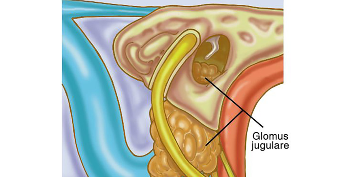
Glomus tumors are tumors from embryonic nerve tissue (embryonic neural crest), which normally form a part of the nervous system known as paragangliomas of the sympathetic nervous system. Glomus tumors are categorized as neuroendocrine neoplasms with their neuro-secreting granules of hormones, which rarely (less than 3 percent) produce enough hormones to affect blood pressure.
The majority (97 percent) of these tumors are benign and are usually small, nourished by very small blood vessels. All these tumors are very vascular, sponge-like filled with blood.
Glomus tumors typically occur in the fourth or fifth decade but can be seen in infancy and as late as the eighth decade. They present more frequently in women.
Symptoms
Glomus tumors can appear with symptoms from affected structures of the ear, including:
- Pulsatile ear noises (tinnitus in synchrony with the heart rate) due to increased blood circulation in the tumor-adjacent to the inner ear
- Hearing loss, from tumor mass interfering with sound transmission or erosion of little bones of hearing or inner ear
- Ear pain
- Hoarseness and swallowing difficulties due to the weakness of nerves to vocal cords and the tongue.
These symptoms vary with the tumor size and location relative to the structures in the ear and skull base.
Treatment
Your BSSNY neurotologist will perform a complete otologic-head and neck examination, including cranial nerve assessment to determine if and which structures may be affected: hearing, facial movement, balance, facial sensitivity to touch, tongue movement, swallowing ability, and vocal cord movement. Your hearing test may include listening to the affected ear by an objective examiner for pulsatile ear noises. Evaluation of the vocal cords will be with fiberoptic laryngeal endoscopy with topical anesthesia (awake), performed in the office as part of a routine initial examination.
Imaging studies such as CT scans and MRI are used along with MRA (angiography of the blood vessels in the head and neck to determine tumor blood supply and extent of tumor).
Management of Glomus tumors is usually with surgical resection in properly selected patients based upon their general health, age, and tumor size. Larger glomus tumors may be preoperatively prepared with embolization to block blood supply to reduce operating time and blood loss. Interventional radiologists skilled in this extremely helpful technique perform embolization.

What’s Going On At BSSNY
-

-

Regional neurosurgery practice can now accept major insurance plans
19 September, 2022 -

Functional neurosurgery brings relief to patients that had given up
29 December, 2021 -

Brain and Spine Surgeons of New York expands practice to Stamford
01 December, 2021